Consultant Orthopaedic & Sports Surgeon
Specialising in minimally invasive arthroscopic procedures and joint replacement surgery for the shoulder, knee, hip, and ankle — helping patients return to the activities they love.
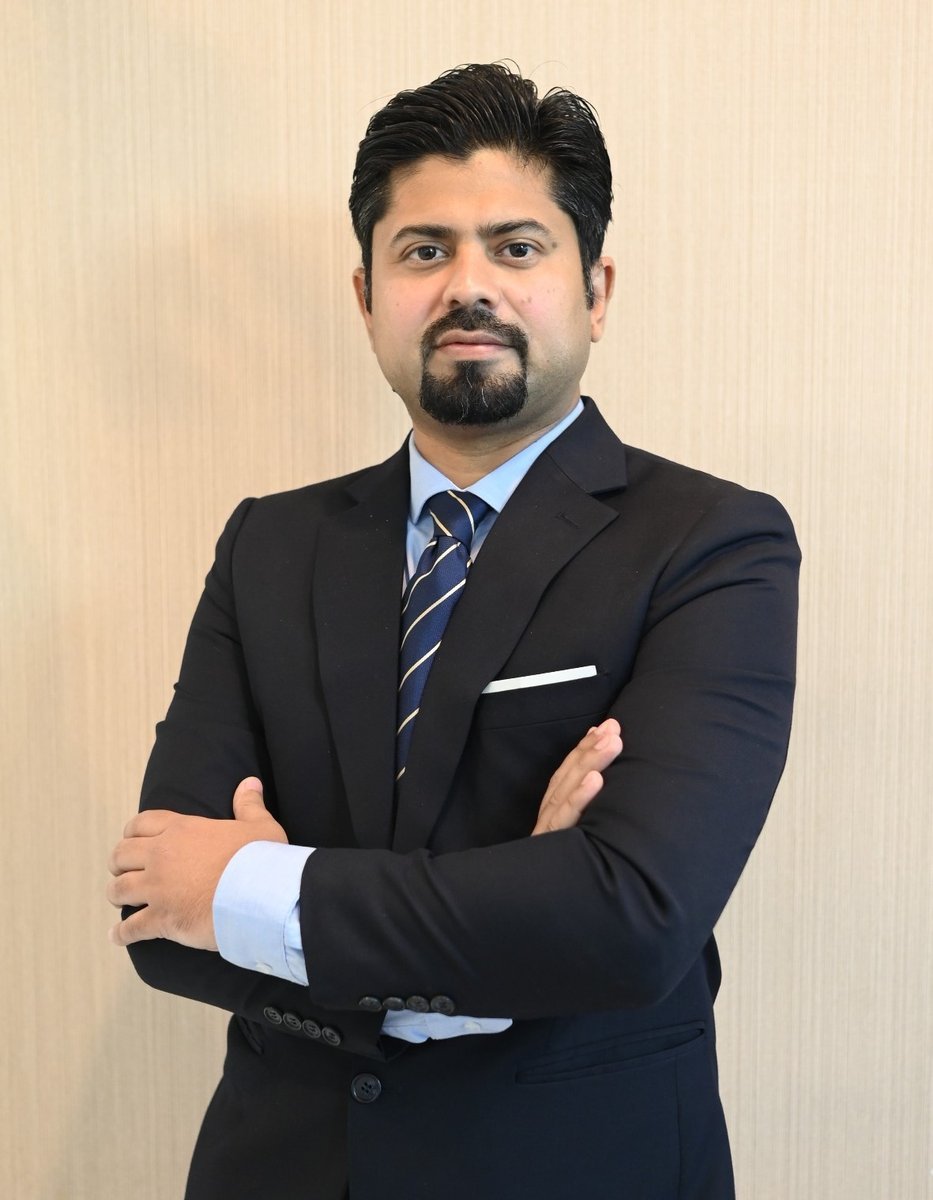
Dr. Nauman Ashraf Malik
MBBS · FIOTA (USA) · FCPS (Tr & Orth) · FSSA
About the Surgeon
With over 15 years of dedicated practice in orthopaedic and sports surgery, Dr. Nauman Ashraf Malik brings exceptional expertise in minimally invasive techniques that reduce recovery times and achieve superior outcomes.
A Fellow of the International Orthopaedic Trauma Association (FIOTA, USA) and the Society of South African Orthopaedic Surgeons (FSSA), Dr. Malik has worked with elite athletes and everyday patients alike — combining cutting-edge surgical technique with a thorough understanding of each patient's goals.
Minimally Invasive Arthroscopy
Keyhole surgery techniques — smaller incisions, less pain, faster return to activity.
Advanced Joint Replacement
Computer-assisted and precision implant placement for longevity and natural function.
Sports-Focused Rehabilitation
Sport-specific recovery programmes designed in collaboration with physiotherapy teams.
Areas of Expertise
From acute sports injuries to complex joint reconstruction — comprehensive orthopaedic care tailored to your goals.
Rotator cuff repair, labral reconstruction, shoulder instability, AC joint injuries, and shoulder replacement.
Arthroscopic · Replacement
ACL/PCL reconstruction, meniscal repair, cartilage restoration, knee replacement, and revision surgery.
Arthroscopic · Replacement
Hip arthroscopy, FAI correction, labral repair, total and partial hip replacement with rapid recovery protocols.
Arthroscopic · Replacement
Ankle arthroscopy, ligament reconstruction, Achilles repair, ankle impingement, and ankle replacement.
Arthroscopic · Reconstruction
Patient Education
Select a topic below to learn about common orthopaedic conditions, their causes, symptoms, and treatments.
The ACL is one of the four main ligaments that stabilise the knee joint, running diagonally through the middle of the knee. It prevents the tibia from sliding out in front of the femur and provides rotational stability.
ACL tears are among the most common sports injuries, affecting athletes who participate in high-demand activities like football, cricket, basketball, and skiing. Approximately 70% of ACL injuries occur through non-contact mechanisms — sudden stopping, pivoting, or landing awkwardly.
Weeks 1–2: Swelling control
RICE protocol, pain management, gentle range of motion exercises begin
Weeks 3–6: Mobility restoration
Full weight-bearing, quad strengthening, proprioception training
Months 2–4: Strength building
Progressive resistance training, neuromuscular control, sport-specific drills begin
Months 6–9: Return to sport
Full running, cutting, pivoting — return to competitive sport after clearance testing
Recovery timelines vary by patient age, fitness level, graft type, and rehabilitation compliance.
The menisci are two C-shaped pieces of fibrocartilage located between the femur and tibia. They act as shock absorbers, distribute load, and enhance joint stability. Each knee has a medial (inner) and lateral (outer) meniscus.
Meniscal tears are extremely common — they can occur due to acute sporting trauma (twisting with the foot planted) or gradually through degenerative wear. Not all tears require surgery; the treatment depends on tear type, location, patient age, and activity demands.
Dr. Malik follows a preservation-first approach — repairing the meniscus wherever blood supply allows. Meniscal preservation reduces the long-term risk of osteoarthritis. When repair is not possible, partial meniscectomy (removing only the damaged tissue) is preferred over total removal.
Arthroscopic suturing of repairable tears in the vascular (red) zone. Best outcomes in young, active patients. Recovery: 3–4 months to sport.
Removal of only the damaged, irreparable fragment. Faster recovery (4–6 weeks) but preserves less tissue than repair.
For patients who have had a previous meniscectomy and are now experiencing pain. A donor allograft replaces the removed meniscus.
Arthroscopic reattachment of the meniscal root using a trans-tibial pull-through technique — critical for restoring hoop tension and preventing rapid cartilage loss.
The PCL is the strongest ligament in the knee, running from the back of the tibia to the front of the femur. It resists forces that would push the tibia backward relative to the femur.
PCL injuries are less common than ACL injuries but are often missed or misdiagnosed. They typically occur from dashboard injuries in motor vehicle accidents, direct blows to the front of the shin, or falling onto a bent knee.
PCL reconstruction uses an anatomic double-bundle technique to restore both the anterolateral and posteromedial bundles of the ligament, providing superior biomechanical outcomes compared to single-bundle techniques.
PCL is twice as strong as the ACL and less commonly torn in isolation
Most PCL injuries also involve other structures — posterolateral corner, MCL, or ACL
Chronic untreated PCL laxity leads to medial and patellofemoral compartment arthritis
The "posterior sag sign" and "posterior drawer test" are key clinical diagnostic tests
MRI is the gold standard imaging study — 99% sensitivity for PCL tears
Full return to sport typically occurs at 9–12 months following PCL reconstruction, after strength testing confirms symmetric limb symmetry index ≥90%.
The rotator cuff is a group of four muscles and their tendons that surround the shoulder joint — supraspinatus, infraspinatus, teres minor, and subscapularis. Together, they keep the humeral head centred in the glenoid socket and power shoulder movement.
Rotator cuff tears are one of the most common causes of shoulder pain and disability. They can result from acute trauma (falling on an outstretched arm) or chronic degeneration from repetitive overhead activity.
Small partial-thickness tears often respond to physiotherapy and corticosteroid injections. Full-thickness tears, especially in active patients under 60, benefit from arthroscopic repair using a double-row technique that restores the tendon's original footprint.
Less than full thickness of the tendon. Conservative treatment often successful (6 weeks physiotherapy then reassess).
Complete disruption of the tendon. Surgical repair recommended for active patients — arthroscopic single or double-row repair.
Multiple tendons involved. Options include tendon transfer, superior capsule reconstruction, or reverse total shoulder replacement.
Osteoarthritis (OA) is the most common form of arthritis, characterised by progressive loss of articular cartilage — the smooth cushioning tissue covering the bone ends in the joint. As cartilage wears away, bone rubs against bone, causing pain, stiffness, and swelling.
Knee OA affects millions worldwide and is a leading cause of disability. While age is the primary risk factor, OA can also develop earlier in life following previous knee injuries — particularly after ligament tears, meniscal damage, or tibial plateau fractures.
For younger patients, Dr. Malik prioritises joint preservation strategies — osteotomy to realign the joint, cartilage restoration procedures, and biological therapies — to delay or avoid the need for total knee replacement.
Weight loss, physiotherapy, activity modification, anti-inflammatory medications, walking aids
Corticosteroid injections for acute flares; hyaluronic acid (viscosupplementation) for joint lubrication
Joint washout, loose body removal, meniscal surgery — limited role in established OA
Tibial or femoral osteotomy to offload the affected compartment — ideal for young, active patients with unicompartmental disease
Partial (unicompartmental) or total knee replacement for end-stage disease not responding to other measures
FAI is a condition where extra bone develops along one or both of the bones that form the hip joint, causing them to fit together improperly. During movement, the irregular surfaces rub against each other and can damage the articular cartilage and labrum.
FAI is increasingly recognised as a major cause of hip pain in young, active adults and athletes. Left untreated, it can cause progressive cartilage damage leading to early-onset hip arthritis.
Hip arthroscopy allows correction of the bony impingement (femoroplasty/acetabuloplasty) and repair of the torn labrum through two or three small portals — avoiding the need for open hip surgery. Most patients return to sport within 4–6 months.
Clinical Assessment
FADIR test (flexion, adduction, internal rotation) — the most sensitive provocative test for FAI
Plain X-Rays
AP pelvis and cross-table lateral views to identify cam or pincer deformity. Alpha angle >55° indicates cam lesion.
MRI Arthrogram
Gadolinium contrast MRI provides detailed assessment of labral tears and articular cartilage damage
Diagnostic Injection
Intra-articular local anaesthetic injection can confirm the hip as the pain source before planning surgery
Common Procedures
Arthroscopic anterior cruciate ligament reconstruction using autograft or allograft — gold standard for return to sport.
Preservation-first approach to meniscal tears — repair where possible, resection only when necessary.
Computer-assisted total knee arthroplasty — precise alignment restores natural biomechanics. Patients walk day one and go home within 2–3 days.
Bone-conserving surgery replacing only the damaged compartment — faster recovery, more natural feel, and easier revision if needed later.
Microfracture, OATS, and osteochondral grafting to restore articular cartilage and delay joint replacement.
Complex ligament reconstruction for posterior cruciate and combined instability patterns.
MPFL reconstruction, tibial tubercle osteotomy, and trochleoplasty for patellar instability and pain.
Minimally Invasive Surgery
Arthroscopic surgery uses a pencil-thin camera and micro-instruments through keyhole incisions — achieving results that previously required large open cuts, with a fraction of the recovery time.
A 4mm fibre-optic camera inserted through a 1cm incision provides a high-definition live view of the joint interior — far superior to MRI for assessing cartilage and soft tissue.
Two 1cm portals give complete access to the knee — the medial and lateral compartments, menisci, cruciate ligaments, and articular cartilage can all be treated without opening the knee.
Rotator cuff repair, labral reconstruction, and impingement release through 2–3 tiny portals — avoiding the large deltoid-splitting incision of traditional open surgery.
Traditional Open Surgery
Arthroscopic Surgery
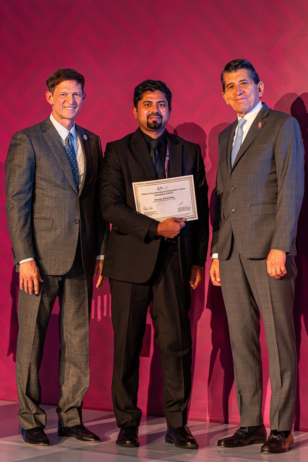
FIOTA — Fellow of the International Orthopaedic Trauma Association
Awarded at the IOTA Annual Congress, USA
International Recognition
Dr. Nauman Ashraf Malik was awarded the Fellowship of the International Orthopaedic Trauma Association (FIOTA) — one of the most prestigious international recognitions in orthopaedic trauma surgery.
The FIOTA designation recognises orthopaedic surgeons who have demonstrated exceptional commitment to advancing the science and practice of trauma surgery on a global scale. The fellowship is awarded at the IOTA Annual Congress and is held by only a select group of surgeons worldwide.
International Orthopaedic Trauma Association (IOTA)
Global body advancing trauma surgery research, education, and clinical practice
Global Standard of Excellence
FIOTA is awarded to surgeons who have made meaningful contributions to orthopaedic trauma at an international level
Pakistan's Representative
One of a small number of Pakistani orthopaedic surgeons to hold the FIOTA fellowship designation
Professional Memberships
Dr. Malik is an active member of the world's most respected orthopaedic and trauma organisations.
Patient Stories
After my ACL reconstruction, I was back on the pitch within 9 months. The level of care and clarity throughout the whole process was exceptional. I felt in safe hands from day one.
Ahmed K.
Football Player · ACL Reconstruction
I had a total knee replacement at 62 and was honestly afraid of the recovery. Dr. Malik explained everything clearly, the surgery went perfectly, and I was walking the very next day. Life without knee pain is extraordinary.
Farrukh K.
Retired Teacher · Total Knee Replacement
My knee replacement was seamless. The pre-op preparation was thorough, and the recovery was faster than I imagined. At 68, I'm now walking five kilometres a day with no pain at all.
Muhammad H.
Retired · Total Knee Replacement
I had a total hip replacement after years of severe arthritis. The minimally invasive approach meant minimal scarring and I was discharged in two days. Three months later I'm hiking again — something I thought I'd never do.
Zainab A.
Teacher · Total Hip Replacement
My hip replacement revision was complicated — the previous implant had failed. Dr. Malik handled it with complete confidence and the result has been outstanding. I'm pain free for the first time in years.
Tariq A.
Engineer · Revision Hip Replacement
The arthroscopic shoulder surgery fixed a rotator cuff tear I'd been living with for two years. I was back to playing squash within six months. The minimally invasive approach made all the difference to my recovery.
Omar M.
Squash Player · Rotator Cuff Repair
Patient Questions
Answers to the questions patients ask most about orthopaedic conditions and surgery.
Not always. Many patients can manage well without surgery, particularly those who are older, less active, or who are willing to modify their activities. However, for younger, active patients — especially those who play pivoting sports — ACL reconstruction is generally recommended to restore full knee stability and reduce the risk of further joint damage. The decision is always made together with the patient, based on their activity goals, knee laxity, and presence of associated injuries such as meniscal tears.
Arthroscopic surgery uses a tiny camera (arthroscope) inserted through small "keyhole" incisions, allowing the surgeon to see and operate on the joint without making a large cut. This results in less tissue damage, reduced post-operative pain, a lower infection risk, and faster recovery compared to traditional open surgery. Dr. Malik performs the vast majority of knee, shoulder, hip, and ankle procedures arthroscopically wherever possible.
Most patients are walking the day after surgery and are discharged home within 2–3 days. By 6 weeks, most patients are walking independently and have stopped using a walking aid. By 3 months, most patients have resumed light activities such as driving and gentle walking. Full recovery — including a return to activities like golf, cycling, and swimming — typically occurs by 6–12 months. The artificial joint typically lasts 15–25 years depending on activity levels and implant type.
It depends on the location and type of tear. The outer third of the meniscus (the "red zone") has a blood supply and can heal with conservative treatment. Tears in this area may resolve with rest, physiotherapy, and time. However, tears in the inner two-thirds (the "white zone") have no blood supply and cannot heal on their own. These may require surgical repair or trimming. Symptoms that persist beyond 6–8 weeks of conservative treatment, or that include locking, catching, or significant instability, generally warrant surgical assessment.
FAI occurs when abnormal bone shapes on the femoral head (cam lesion) or acetabulum (pincer lesion) cause them to rub together during movement, damaging the labrum and articular cartilage. It commonly presents as groin pain in young, active adults, particularly with hip flexion activities. Treatment starts conservatively with physiotherapy and activity modification. If pain persists, hip arthroscopy allows correction of the bony deformity and repair of any labral tears, with most patients returning to sport within 4–6 months.
A second opinion is always reasonable — particularly when: (1) you have been recommended a major surgery and want to confirm it's necessary; (2) your symptoms are not improving despite treatment; (3) you have a complex or unusual diagnosis; or (4) you are considering a high-risk procedure with significant recovery demands. Dr. Malik welcomes patients seeking a second opinion and offers thorough MRI/X-ray review consultations for patients travelling from outside Islamabad.
Please bring: any previous X-rays, MRI or CT scan reports and imaging CDs/films; a list of current medications and allergies; a summary from your referring doctor if available; your health insurance details; and a list of questions you'd like answered. Wearing or bringing comfortable shorts is helpful for lower limb examinations. If you have had previous surgery on the affected joint, please bring the operative report if possible.
Arthroscopic rotator cuff repair typically takes 1–2 hours under general anaesthesia. Most patients go home the same day. Recovery involves 4–6 weeks in a sling to protect the repair, followed by progressive physiotherapy. Gentle range of motion begins at 6 weeks, strengthening at 3 months, and full overhead activity at 6 months. Return to sport or heavy labour typically takes 9–12 months. Larger tears take longer to heal than smaller ones.
Get in Touch
Take the first step toward pain-free movement. Consultations available at two clinics in Islamabad.
Ali Medical Center
Kohistan Road, F-8 Markaz, Islamabad
Mon–Fri: 2:00 PM – 9:00 PM · Sat: 5:00 PM – 9:00 PM
Enfield Royal Clinics
Ammar Plaza, F-7 Markaz, Islamabad
Mon–Fri: 12:00 PM – 2:00 PM
Phone
051-8090200
051-2656411 / 2656412 / 2656413
We will get back to you within one working day.